|
|
|
Technical data Histology of domestic animals for veterinary medicine part I, 24 prepared microscope slidesArticle no: LIE-84000

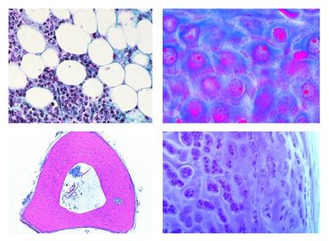
Function and Applications Histology of domestic animals for veterinary medicine, part I, 24 prepared microscope slides - - Simple columnar epithelium, in t.s. of small intestine of pig - Pseudostratified ciliated columnar epithelium, in t.s. of trachea - White fibrous tissue, l.s. of tendon of cow - Yellow elastic cartilage, ear of rabbit or pig, t.s. - Bone development, intracartilaginous ossification in foetal finger or toe, l.s. - Striated (skeletal) muscle of cat l.s. - Heart muscle of mammal, l.s. and t.s. - Heart of mouse, entire sagittal l.s. - Trachea of cat or rabbit, l.s. - Motor nerve cells, smear preparation from spinal cord of ox stained for Nissl bodies - Spleen of rabbit, t.s. showing capsula, pulp etc. - Lymph node of pig, t.s. routine stained - Adrenal gland (Gl. suprarenalis) of rabbit, t.s. through cortex and medulla - Thyroid gland of cow, sec. showing colloid - Thymus of young calf, t.s. with Hassall bodies - Adipose tissue of pig, section fat removed to show the cells - Oesophagus of rabbit or dog, t.s. - Rumen of cow, t.s. - Reticulum of cow, t.s. - Omasum of cow, t.s. - Abomasum of cow, t.s. - Vermiform appendix, rabbit t.s. - Colon of pig, t.s. stained with muci-carmine or PAS for demonstration of mucous cells - Ureter of pig, t.s. The microslides are supplied in a slide box. |
|
|
Robert-Bosch-Breite 10 – 37079 Göttingen – Germany
www.phywe.com

