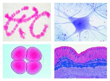
Technical data School Set D for General Biology, Supplementary Set, 50 microscope slidesArticle no: LIE-750  Function and Applications No. 750 School Set D for General Biology, Supplementary Set 50 microscope slides - - Histology of Man and Mammals - Ciliated epithelium, in t.s. of fallopian tube of pig - Tendon of cow, l.s. showing white fibrous tissue, stained for fibres and cells - Heart muscle, human, t.s. and l.s., branched fibres with central nuclei and intercalated discs - Lymph gland of pig, t.s. showing lymphoid tissue - Esophagus of cat, t.s. with stratified squamous epithelium, muscular layers - Stomach of cat, t.s. through fundic region showing gastric glands - Large intestine (colon), t.s. special stained for the mucous cells - Pancreas of pig, sec. showing islets of Langerhans - Thyroid gland of pig, sec. showing glandular epithelium and colloid - Adrenal gland of cat, t.s. through cortex and medulla - Sperm of bull (spermatozoa), smear - Motor nerve cells, smear from spinal cord of cow showing w.m. of motor nerve cells and their processes - Cerebrum, human, t.s. of cortex showing pyramidal cells and fibrous region - Human skin from palm, v.s. showing cornified epidermis, germinative zone, sweat glands - Zoology - Distomum hepaticum (Fasciola), the beef liver fluke, w.m. and stained for general study of internal organs - Taenia spec., tapeworm, w.m. of mature proglottids - Culex pipiens, mosquito, head and piercing-sucking mouth parts of female, w.m. - Culex pipiens, mosquito, head and reduced mouth parts of male, w.m. - Cimex lectularius, bed bug, w.m. of adult specimen - Cytology and Genetics - Mitochondria, in thin sec. through liver or kidney, special staining technique - Golgi apparatus, t.s. through spinal ganglion, special staining technique - Chloroplasts, in leaf of Elodea or Mnium, special stained - Aleurone grains, in sec. of Ricinus endosperm - Storage, section of liver or kidney, vital stained with trypan-blue to demonstrate storage in epithelial cells - DNA in cell nuclei, demonstrated by Feulgen staining technique - DNA and RNA, fixed and stained with methyl green and pyronine to show DNA and RNA in different colors - Giant chromosomes from the salivary gland of Chironomus. Individual genes and puffs can be observed - Human chromosomes, spread in the stage of metaphase, for counting chromosomes - Meiotic and mitotic stages in sec. of crayfish testis. Nuclear spindles are present - Maturation divisions in ova of Ascaris megalocephala, different stages, iron-hematoxylin stained - Cleavage stages in ova of Ascaris megalocephala, iron-hematoxylin stained - Bacteria and Diseased Organs of Man - Escherichia coli, bacteria from colon, probably pathogenic, smear Gram stained - Eberthella typhi, causing typhoid fever, smear from culture, Gram stained - Tuberculous lung, t.s. of diseased human lung showing miliary tubercles in tissue - Coal dust lung (Anthracosis pulmonum), t.s. of human smoker’s lung - Liver cirrhosis of man caused by alcohol abuse, t.s. showing degeneration of liver cells - Arteriosclerosis, t.s. of diseased human coronary artery showing sclerotic changes in the arterial wall - Metastatic carcinoma (cancer) of human liver, t.s. - Embryology - Sea-urchin development (Psammechinus miliaris), composite slide with two cell, four cell and eight cell diseased human coronary artery showing sclerotic changes in the arterial wall - Metastatic carcinoma (cancer) of human liver, t.s. - Embryology - Sea-urchin development (Psammechinus miliaris), composite slide with two cell, four cell and eight cell The microslides are supplied in a slide box. |
Robert-Bosch-Breite 10 – 37079 Göttingen – Germany
www.phywe.com

