Principle
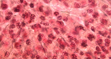
The nucleus is recognizable under the light microscope as a circular object. It can be seen even without previous staining. The nucleus is the control center of many cellular processes and harbors the hereditary information. The nucleus contains filamentous structures (chromatin) which after staining appear as a homogenous mass. Cell division always commences with a division of the nucleus (mitosis). In preparation to this division, the filaments retract and become shorter and thicker. Staining now makes distinctive objects visible, i.e. the chromosomes. The genetic information they contain has already undergone reduplication. The membrane that envelopes the nucleus dissolves, the chromosomes gather in the center of the cell. Attached to the spindle apparatus they migrate to the poles of the cell, where they form two new nuclei. Only then does the body of the cell divide and thus two daughter cells are created.
Benefits
- Experiment is part of a complete solution set with a total of 50 experiments for all microscopy applications
- With student worksheet, appropriate for all class levels
- With detailed instructor information, incl. sample microscopy image
- Optimized for tight schedules, i.e. minimum preparation time required
- Microscopy solution set specifically designed to include all required accessories
- Content available with matching multimedia files
Tasks
Study plant cells undergoing mitosis under the microscope!
Learning objectives
- Nucleus
- Chromosomes
- Cell division
- Chromatin


